
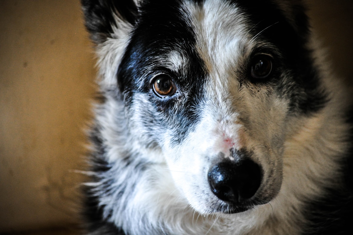
There is an exquisite intimacy in looking into the eyes of a beloved canine companion. In those warm, expressive pools of amber, hazel, or deep brown, we read an open book of unfiltered emotion: unyielding loyalty, boundless joy, gentle empathy, and quiet anticipation. Yet, because we gaze into our dogs’ eyes so frequently during daily moments of affection, subtle physical alterations often develop in plain sight before our conscious minds register the danger. A slight milky haze across the pupil, a fine network of red spider veins across the sclera, a subtle squint when stepping into bright morning sunlight, or a crusty bead of dried discharge at the inner corner is easily dismissed as natural aging or minor dust irritation.
In small animal clinical practice, there is no medical discipline where hesitation or delay is more catastrophic than veterinary ophthalmology. The eye is not merely another organ; it is a delicate, pressurized, highly specialized optical sphere where microscopic cellular injuries can escalate into permanent, irreversible blindness within twenty-four to forty-eight hours. A minor superficial scratch on the cornea can be colonized by collagen-destroying bacteria, transforming into a melting ulcer that ruptures the globe overnight. Similarly, an acute spike in intraocular pressure from canine glaucoma can permanently destroy every retinal ganglion cell in the back of the eye in less than twelve hours, extinguishing vision forever while causing agonizing, silent migraine-like pain.
According to clinical consensus guidelines published by the American Veterinary Medical Association (AVMA) and health resources from the American Kennel Club (AKC), early detection of ocular changes is the single most important factor determining whether an injured or diseased eye can be saved. Protecting your dog’s sight requires knowing how to read the visual language of the canine eye, understanding the anatomical structures that govern ocular health, distinguishing harmless age-related changes from sight-threatening emergencies, and recognizing the precise red-flag symptoms that demand immediate, same-day hospital intervention.
Anatomical Architecture of the Specialized Canine Eye
To understand how ocular diseases manifest and progress, one must first explore the unique structural architecture of the canine eye. The outer protective tunic of the globe is composed of two continuous fibrous structures: the posterior opaque white sclera, and the anterior transparent, dome-shaped cornea.
The cornea is a living optical marvel, measuring less than one millimeter in thickness across most canine breeds, yet composed of four distinct anatomical layers:
- Outermost Stratified Squamous Epithelium: A five-to-seven-cell-layer hydrophobic barrier that prevents tear fluid and environmental bacteria from invading the underlying stroma.
- Corneal Stroma: Comprising ninety percent of total corneal thickness, the stroma consists of precisely spaced, parallel lamellae of Type I collagen fibers.
- Descemet’s Membrane: A resilient, elastic basement membrane secreted by the underlying endothelial monolayer, acting as the final physical barrier against corneal rupture.
- Innermost Endothelial Monolayer: A single layer of specialized non-regenerative hexagonal cells equipped with metabolic sodium-potassium ATPase pumps. These pumps constantly pump fluid out of the stroma back into the anterior chamber, maintaining a state of relative dehydration: termed corneal deturgescence: that is strictly mandatory for optical transparency.
The Aqueous Fluid Dynamics and Drainage Angle
Directly behind the cornea lies the anterior chamber, filled with clear, nutrient-rich aqueous humor fluid produced continuously by the ciliary body epithelium. Aqueous humor fulfills the role of blood for the avascular cornea and lens, circulating forward through the pupil before draining out of the eye through a microscopic sieve-like structure at the junction of the iris and cornea known as the iridocorneal drainage angle (pectinate ligaments). In a healthy eye, fluid production matches fluid drainage in perfect equilibrium, maintaining a stable intraocular pressure (IOP) between 15 and 25 millimeters of mercury (mmHg).
The Crystalline Lens and Tapetum Lucidum
Positioned immediately behind the colored iris is the biconvex crystalline lens, suspended by delicate ciliary zonules. The lens fine-tunes light refraction, focusing optical images across the vast vitreous chamber onto the neural retina lining the back of the globe. Positioned directly behind the retina is an evolutionary marvel called the tapetum lucidum: a cellular reflective mirror that bounces unabsorbed photons back through the photoreceptors a second time, providing canines with superior twilight and nocturnal vision while generating the brilliant green, yellow, or blue eye shine seen when light hits a dog’s eyes in the dark.
The Nictitating Membrane (Third Eyelid)
Canines possess a specialized anatomical structure absent in human primates: the third eyelid, clinically designated as the nictitating membrane. Tucked quietly into the inner corner (medial canthus) of each eye, the third eyelid is a T-shaped cartilage flap covered in conjunctival mucous membrane. Within its base resides the superficial gland of the third eyelid, responsible for producing thirty to fifty percent of the aqueous portion of the precorneal tear film. Every time a dog blinks, the third eyelid sweeps passively across the cornea like an internal windshield wiper, clearing particulate debris and distributing protective immunoglobulins and lubricating tears.
Decoding Critical Color Changes in Your Dog’s Eyes
Color changes within the ocular structures are among the earliest and most unmistakable warning signs that an active disease process is underway.
1. The Red Eye: Conjunctival Hyperemia versus Episcleral Injection
Redness in the white of the eye is one of the most common presenting complaints in veterinary clinics. However, clinicians must distinguish between two completely different vascular networks:
- Superficial Conjunctival Hyperemia: Fine, branching, bright-red blood vessels located on the mobile conjunctival membrane that covers the sclera. These vessels move freely when the eyelid is gently shifted and blanch immediately when a drop of topical phenylephrine is applied. Conjunctival redness is typical of surface irritation, conjunctivitis, dry eye, or mild environmental allergies.
- Deep Episcleral / Ciliary Injection: Straight, engorged, dark-red or purple blood vessels deeply anchored into the rigid white sclera. These vessels radiate outward from the corneal margin like spokes on a bicycle wheel and do not move when the eyelid is manipulated. Deep episcleral injection is a clinical red flag signaling severe, sight-threatening intraocular disease: specifically acute glaucoma or anterior uveitis.
2. The Cloudy Eye: Nuclear Sclerosis versus Cataracts
One of the most frequent diagnostic dilemmas faced by pet owners is distinguishing between benign age-related nuclear sclerosis and sight-threatening cataracts in middle-aged and senior dogs:
- Nuclear Sclerosis (Lenticular Sclerosis): A completely normal, benign physiological consequence of aging seen in virtually all dogs over seven years old. As the dog ages, the lens continuously produces new fiber cells from its outer cortex. Because the rigid lens capsule cannot expand, older fibers are compressed tightly inward into the central nucleus. This optical compaction reflects light, creating a hazy, pearlescent grayish-blue marble appearance deep within the pupil. Crucially, nuclear sclerosis does not block light transmission significantly; light passes freely through to the retina, allowing the dog to maintain functional vision, and ophthalmoscopic examination reveals an unobstructed view of the tapetal fundus. It requires zero medical intervention.
- True Canine Cataracts: A true cataract is a pathological, irreversible opacification of the lens fibers or lens capsule. Cataracts physically block light from reaching the retina, resulting in progressive visual impairment, dilated pupils, bumping into furniture, and eventual total blindness. Cataracts can arise from genetic inheritance, diabetes mellitus (where high glucose levels convert into sorbitol, pulling water into the lens), blunt trauma, or chronic intraocular inflammation. Furthermore, mature cataracts can leak lens proteins into the eye, inducing painful lens-induced uveitis and secondary glaucoma, requiring surgical removal via phacoemulsification.
3. The Blue Eye: Corneal Edema
Unlike cataracts or nuclear sclerosis, which reside deep inside the pupil within the lens, corneal edema occurs directly within the cornea itself, transforming the crystal-clear optical window into a milky, ground-glass, opaque light-blue surface:
- Loss of Deturgescence: Corneal edema occurs when fluid floods the corneal stroma, separating collagen lamellae and scattering light waves.
- Epithelial Breach: A deep corneal ulcer allows precorneal tear fluid to penetrate directly into the stroma from the outside.
- Endothelial Pump Failure: Endothelial dystrophy, severe anterior uveitis, or skyrocketing intraocular pressure in acute glaucoma destroys the endothelial fluid pumps, allowing aqueous humor to saturate the cornea from the inside.
4. Hyphema and Hypopyon: Blood and Pus in the Anterior Chamber
The appearance of opaque fluids settling in the anterior chamber between the cornea and iris represents an alarming clinical presentation:
- Hyphema: The accumulation of free liquid red blood in the anterior chamber, pooling at the bottom of the eye or filling the entire eye with dark crimson fluid. Hyphema signals severe blunt trauma, intraocular vascular tumors (melanoma, ciliary adenocarcinoma, lymphoma), systemic arterial hypertension, retinal detachment, or fatal systemic coagulopathies like rat poison ingestion.
- Hypopyon: The settling of dense, creamy-white or yellowish purulent exudate (white blood cells and fibrin) in the anterior chamber, indicating severe intraocular bacterial infection (endophthalmitis) or aggressive immune-mediated uveitis.
Specialized diagnostic protocols published by the American College of Veterinary Ophthalmologists (ACVO) emphasize immediate examination whenever corneal integrity or intraocular pressures deviate from normal ranges.
Pupil Abnormalities and Neurological Asymmetry (Anisocoria)
Pupil size, shape, and reactivity provide direct windows into both ocular health and central nervous system integrity.
Evaluating Anisocoria (Unequal Pupils)
Anisocoria is the presence of asymmetrical pupil sizes where one pupil is abnormally constricted and the other is abnormally dilated. Determining which eye is the abnormal one requires testing pupillary light reflexes (PLR) in both bright light and complete darkness:
- Constricted Pupil (Miosis) Pathologies: If the smaller pupil is the diseased eye, it frequently indicates acute Anterior Uveitis (where prostaglandin release causes intense ciliary muscle spasm) or Horner’s Syndrome. Horner’s syndrome is a neurological disorder resulting from damage to the sympathetic nerve pathway, presenting as miosis, upper eyelid drooping (ptosis), a sunken eyeball (enophthalmos), and prominent elevation of the third eyelid.
- Dilated Pupil (Mydriasis) Pathologies: If the larger pupil is the diseased eye and fails to constrict in response to bright light, this is an emergency of the highest order: Acute Canine Glaucoma. Extreme intraocular pressure paralyzes the iris sphincter muscle, locking the pupil in a wide, unresponsive dilation while cutting off blood flow to the optic nerve.
Corneal Ulcers: From Superficial Scratches to Melting Disasters
The cornea is one of the most densely innervated tissues in the animal body, packed with thousands of sensory nerve endings per square millimeter. Consequently, even microscopic corneal abrasions cause intense pain.
The Spectrum of Corneal Ulceration
- Superficial Corneal Ulcers: A simple loss of the outer epithelial layer, commonly caused by a scrape from a thorn, cat scratch, or foreign body. With appropriate topical antibiotic therapy to prevent infection, healthy basal epithelial cells slide and multiply, healing the defect completely within three to five days without scarring.
- Indolent (Boxer) Ulcers / Spontaneous Chronic Corneal Epithelial Defects (SCCEDs): A non-healing ulcer seen predominantly in Boxers, Golden Retrievers, and middle-aged dogs. The underlying corneal stroma develops a defective basement membrane, preventing newly formed epithelial cells from adhering securely. The loose epithelial edges roll up, and the ulcer persists for weeks or months. Treatment requires debridement and diamond-burr burring or anterior stromal puncture (keratotomy) to allow new cells to anchor.
- Melting Corneal Ulcers (Keratomalacia): The most terrifying corneal emergency in veterinary medicine. When a corneal ulcer becomes infected with virulent bacteria: particularly Pseudomonas aeruginosa or beta-hemolytic Streptococcus: the bacteria and dying host white blood cells release massive quantities of collagenases, gelatinases, and matrix metalloproteinases. These enzymes literally dissolve and digest the corneal stroma. The cornea transforms into a soft, grayish, gelatinous liquefactive mush. A melting ulcer can dissolve completely through the stroma in less than twelve hours, leading to descemetocele formation, full corneal perforation, iris prolapse, and complete loss of the eye.
Clinical standards established by the American Animal Hospital Association (AAHA) emphasize the necessity of structured diagnostic evaluations and routine monitoring protocols.
Keratoconjunctivitis Sicca (KCS): The Agony of Dry Eye
Keratoconjunctivitis Sicca, universally known as canine dry eye, is a chronic, painful ophthalmic condition characterized by severe deficiency in the aqueous tear film produced by the lacrimal and third eyelid glands.
The Tri-Layered Tear Film and Immune-Mediated Destruction
The canine precorneal tear film is composed of three distinct layers:
- Inner Mucin Layer: Secreted by conjunctival goblet cells, coating the hydrophobic corneal epithelium so tears can spread evenly.
- Middle Aqueous Layer: Secreted by the orbital lacrimal gland and the gland of the third eyelid, providing oxygen, nutrients, antibacterial lysozyme, and moisture.
- Outer Lipid Layer: Secreted by the Meibomian glands along the eyelid margins, preventing evaporation of the underlying watery layer.
In over seventy-five percent of canine KCS cases, the underlying cause is autoimmune, T-cell-mediated destruction of the lacrimal glandular tissue. Breeds heavily predisposed include the Cavalier King Charles Spaniel, English Bulldog, Pug, West Highland White Terrier, and Shih Tzu. As aqueous tear production drops from normal levels (greater than 15 mm/minute on a Schirmer Tear Test) down to single digits or zero, the eye dries out. Desiccated goblet cells produce massive amounts of thick, sticky, ropy mucopurulent yellow discharge that coats the eye like glue. Without lubrication, chronic friction causes the cornea to turn black with scar tissue and pigment (pigmentary keratitis), leading to complete blindness.
Prolapsed Gland of the Third Eyelid (Cherry Eye)
Cherry Eye is the colloquial term for the prolapse of the superficial gland of the nictitating membrane. In young dogs: particularly brachycephalic breeds like French Bulldogs, English Bulldogs, Cocker Spaniels, and Beagles: the fibrous connective tissue retinaculum that anchors the tear gland to the periorbital tissue is congenitally weak or absent.
The gland pops out of its normal anatomical pocket behind the third eyelid, swelling into a bright pink, fleshy, oval mass protruding from the medial corner of the eye. While cherry eye looks alarming, it is not an acute emergency; however, it must never be ignored. In historical decades, veterinarians frequently removed the prolapsed gland surgically. Today, this practice is recognized as medical malpractice: excising the gland eliminates up to fifty percent of the eye’s aqueous tear production, virtually guaranteeing that the dog will develop severe, blinding Keratoconjunctivitis Sicca later in life. Modern ophthalmic surgery utilizes mucosal pocketing techniques (such as the Morgan pocket procedure) that tuck the gland back into its normal anatomical position, preserving vital tear production for life.
According to clinical pathology references published in the Merck Veterinary Manual, early identification of underlying physiological changes provides the highest probability of positive long-term therapeutic outcomes.
The Essential Veterinary Ophthalmic Examination Protocol
When presenting your dog for an eye evaluation, your veterinarian will utilize three indispensable diagnostic tests before ever instilling therapeutic medications:
1. The Schirmer Tear Test (STT)
A specialized sterile paper strip with a calibrated millimeter scale is placed inside the lower eyelid conjunctival sac for exactly sixty seconds. Capillary action draws tears down the strip. A normal reading is between 15 and 25 millimeters of wetting per minute. Readings below 10 mm/min confirm clinical dry eye, while readings above 25 mm/min indicate excessive reflex tearing from painful irritation.
2. Fluorescein Dye Corneal Staining
Fluorescein is a water-soluble orange dye that fluoresces brilliant emerald green under cobalt blue light. Intact corneal epithelium is hydrophobic and completely repels the dye. However, if the epithelium is scratched, ulcerated, or ruptured, the underlying hydrophilic stroma absorbs the dye like a sponge. Any green fluorescence confirms a corneal ulcer, establishing its exact depth, surface area, and border configuration. Furthermore, fluorescein dye flowing through the nasolacrimal duct into the nostrils (Jones test) confirms normal tear drainage passage.
3. Rebound or Applanation Tonometry
Using precision veterinary tonometers (such as the TonoVet or Tono-Pen), the clinician measures intraocular pressure (IOP) in millimeters of mercury. Normal IOP is 15 to 25 mmHg. An IOP above 25 to 30 mmHg confirms glaucoma, while an IOP below 10 mmHg confirms active anterior uveitis.
The Deadly Hazard of Using Leftover Eye Drops
One of the most catastrophic mistakes dog owners make is reaching into their medicine cabinet to use leftover prescription eye drops: or human over-the-counter drops like Visine: when their dog’s eye appears red or irritated.
Many veterinary combination eye drops (such as NeoPolyDex) contain potent corticosteroids like dexamethasone or prednisolone. If a dog has a corneal scratch or ulcer, applying a steroid eye drop is disastrous: corticosteroids paralyze white blood cell defense, stimulate collagen-destroying enzymes, and turn a simple microscopic scratch into a ruptured melting ulcer within forty-eight hours. Never apply any eye drop containing a steroid to a canine eye without a negative fluorescein stain performed that same day by a veterinarian.
Eyelid Conformation Disorders: Entropion, Ectropion, and Distichiasis
The eyelids and eyelashes serve as the first physical line of defense for the ocular globe. However, congenital anatomical malformations can transform these protective structures into instruments of chronic corneal trauma.
1. Entropion: The Inward Rolling Eyelid
Entropion is the inward inversion of the eyelid margin, causing the hair-bearing outer skin, stiff facial fur, and eyelashes to rub directly against the sensitive cornea with every blink:
- Breeds Heavily Affected: Shar-Peis, English Bulldogs, Chow Chows, Rottweilers, and Golden Retrievers.
- Pathological Consequences: Relentless trichiasis (hair friction) creates painful corneal abrasions, severe blepharospasm, reflex lacrimation, and progressive vascularization. If uncorrected, the chronic abrasion develops into a non-healing corneal ulcer or dense black pigmentary keratitis.
- Surgical Resolution: Corrected through the Hotz-Celsus procedure, an ophthalmic blepharoplasty where a crescent-shaped strip of skin and orbicularis oculi muscle is excised beneath the inverted lid, rolling the eyelid margin back into perfect anatomical alignment.
2. Ectropion: The Slack Outward Eversion
Ectropion is the outward sagging or eversion of the lower eyelid margin, exposing the pale pink palpebral conjunctiva to the open environment:
- Breeds Affected: Basset Hounds, Bloodhounds, Saint Bernards, and Newfoundlands.
- Clinical Signs: Environmental wind, pollen, and dust settle into the exposed conjunctival cul-de-sac, causing chronic follicular conjunctivitis, brown tear staining, and persistent mucous discharge.
3. Distichiasis and Ectopic Cilia: Rogue Eyelashes
In distichiasis, abnormal extra eyelashes emerge from the Meibomian gland orifices along the eyelid margin. If the hairs are fine and silky, they float harmlessly in the tear film; however, if they are stiff, they scratch the cornea like wire brushes. Even more destructive are Ectopic Cilia: eyelashes that emerge through the palpebral conjunctiva on the inner surface of the upper eyelid, pointing directly perpendicular into the cornea. Ectopic cilia cause intense, excruciating focal ulcers in young dogs and require surgical excision under an operating microscope.
Retinal Emergencies: Progressive Retinal Atrophy and SARDS
While corneal and lens diseases alter the front of the eye, catastrophic pathologies within the neural retina extinguish vision at the neuro-sensory level.
1. Progressive Retinal Atrophy (PRA)
Progressive Retinal Atrophy encompasses a family of inherited genetic mutations causing bilateral, symmetrical photoreceptor degeneration:
- Nyctalopia (Night Blindness): Rod photoreceptors, responsible for dim-light vision, degenerate first. Owners notice their dog hesitating to enter dark hallways, bumping into objects in the backyard at night, or refusing to go outside after sunset.
- Progressive Diurnal Vision Loss: Over several months to years, cone photoreceptors degenerate as well, resulting in total day blindness. The pupils become widely dilated, sluggishly responsive to light, and the tapetal fundus develops hyper-reflective golden sheen on ophthalmoscopic exam. Genetic DNA panel testing allows responsible breeders to screen for carrier mutations before breeding.
2. Sudden Acquired Retinal Degeneration Syndrome (SARDS)
SARDS is one of the most heartbreaking and mysterious ophthalmic conditions in veterinary medicine, characterized by rapid, complete, irreversible death of all retinal photoreceptors over a window of twenty-four to seventy-two hours:
- Patient Demographics: Most commonly strikes middle-aged (7 to 10 years old), slightly overweight spayed female canines, particularly Dachshunds, Miniature Schnauzers, and mixed-breed dogs.
- Concurrent Endocrine Signs: Dogs with SARDS frequently display acute polydipsia, polyuria, polyphagia, and sudden weight gain mirroring Cushing’s disease weeks before blindness strikes.
- The Diagnostic Paradox: When an owner rushes their suddenly blind dog to a clinic, the eyes appear completely normal on visual exam: the corneas are clear, the lenses are transparent, and the ophthalmoscopic retinal fundus looks pristine. Confirmatory diagnosis requires an Electroretinogram (ERG): in SARDS, the ERG displays a completely flat electrical line, confirming total loss of retinal photoreceptor function.
Systemic Vascular and Metabolic Diseases Manifesting in the Eye
The eye is uniquely transparent, allowing clinicians to directly observe living microvascular circulation without surgery. Systemic diseases frequently declare themselves in the canine eye before causing clinical collapse elsewhere in the body:
- Systemic Arterial Hypertension: In dogs suffering from chronic kidney disease or pheochromocytoma, high blood pressure ruptures delicate retinal capillaries, creating flame-shaped retinal hemorrhages, bullous retinal detachment, and sudden hyphema.
- Diabetic Cataract Genesis: Within twelve months of diagnosis, over seventy-five percent of diabetic dogs develop bilateral mature cataracts. High circulating glucose enters the lens and saturates the hexokinase pathway, shunting glucose into the sorbitol pathway. Intracellular sorbitol creates massive hyperosmotic swelling, rupturing lens fibers within days.
- Lymphoma and Systemic Neoplasia: Malignant lymphoblasts frequently infiltrate the highly vascular uveal tract, causing severe bilateral anterior uveitis, hypopyon, and secondary glaucoma as the initial presenting sign of multicentric lymphoma.
First Aid for Ocular Trauma: Emergency Field Interventions
When sudden physical trauma strikes your dog’s eye, administering correct, calm emergency first aid at home before transport to the veterinary hospital can save your companion’s vision:
Critical First Aid Rules for Ocular Emergencies
- Chemical or Irritant Contamination: If household cleaning chemicals, lawn sprays, or pool shock splash into your dog’s eye, flush the eye immediately with sterile saline eyewash (or clean lukewarm tap water) continuously for fifteen to twenty minutes. Hold the eyelids open gently and direct the stream from the inner corner outward to dilute caustic agents.
- Traumatic Proptosis (Eye Popped from Socket): In brachycephalic dogs with shallow orbits (Pugs, French Bulldogs, Shih Tzus), blunt head trauma or rough scruff handling can cause the eyeball to pop forward beyond the eyelids. Never attempt to push the eyeball back into the socket yourself. Immediately soak a clean gauze pad or washcloth in sterile saline (or clean warm water) and gently drape it over the protruding eye to keep tissues moist. Transport the dog to an emergency surgical facility immediately; surgical repositioning within one hour offers the best chance of salvaging the globe and vision.
- Penetrating Foreign Objects: If a thorn, splinter, or wire has pierced the cornea, do not attempt to pull it out. Removing the object can cause immediate collapse of the anterior chamber and catastrophic iris prolapse. Place an Elizabethan cone collar on your dog immediately to prevent pawing, and transport them with minimal head movement.
Canine Ocular Tonometry and Slit-Lamp Biomicroscopy Diagnostic Protocols
Navigating canine health challenges requires a systematic diagnostic methodology grounded in evidence-based veterinary pathology. Whether evaluating insidious systemic complaints such as mild polydipsia, post-rest lameness, or subtle ocular alterations, clinical specialists emphasize the necessity of combining objective laboratory biomarkers with detailed longitudinal symptom tracking. Routine baseline screening – encompassing complete blood counts, comprehensive biochemical profiles, high-resolution digital imaging, and specific serological antibody assays – enables veterinary teams to intercept metabolic and degenerative disorders during subclinical stages.
Corneal Endothelial Decompensation and Microvascular Anterior Chamber Flare
Systemic illness in canines frequently manifests through alterations in microvascular capillary perfusion and endothelial barrier permeability. Inflammatory cytokines released during infectious or autoimmune states stimulate endothelial cell activation, promoting localized vascular stasis, plasma extravasation into interstitial spaces, and micro-thrombosis. Monitoring capillary refill time, serum lactate levels, and base excess provides clinicians with real-time metrics of tissue oxygenation and hemodynamic stability, guiding targeted fluid therapy and pharmacological interventions before organ dysfunction becomes irreversible.
Immune-Mediated Uveitis and Secondary Glaucoma Pathophysiological Cascades
The gut-associated lymphoid tissue (GALT) plays a central role in maintaining systemic immune tolerance while protecting against enteric pathogens. Disruptions in mucosal tight junctions (colloquially termed intestinal permeability) permit dietary antigens and bacterial lipopolysaccharides to enter the lamina propria, triggering chronic low-grade inflammation that can manifest as intermittent gastrointestinal borborygmi, selective appetite loss, or distant dermatological flare-ups. Implementing targeted hypoallergenic diets, mucosal cytoprotectants, and scientifically validated synbiotic therapy restores mucosal barrier integrity and stabilizes immune equilibrium.
Geriatric Ophthalmic Care: Distinguishing Nuclear Sclerosis from Cataractogenesis
Optimizing senior canine longevity requires a multi-modal wellness approach that addresses cognitive preservation, joint comfort, and metabolic stability. Incorporating omega-3 polyunsaturated fatty acids (EPA and DHA), multimodal analgesia protocols (including modern anti-NGF monoclonal antibodies), and gentle low-impact physical exercise maintains lean muscle mass and cognitive engagement. Regular veterinary health evaluations every six months ensure that age-related degenerative changes are managed proactively, preserving dignity and comfort throughout the golden years.
Emergency Veterinary Referral Timelines for Acute Vision-Threatening Ocular Signs
Maintaining long-term wellness for companion dogs requires an active, ongoing partnership between dedicated guardians and qualified veterinary professionals. Establishing regular six-month wellness examinations, conducting routine diagnostic laboratory panels, and maintaining longitudinal records of behavioral patterns and dietary responses provides clinicians with essential baseline metrics. By adopting an evidence-based preventative mindset and addressing subtle environmental and physical changes promptly, pet parents ensure their canine companions enjoy optimal vitality, emotional comfort, and flourishing health throughout every developmental life stage.
Comparative Diagnostic Matrix: Major Canine Ocular Conditions
To assist dog owners and clinicians in evaluating ocular presentations and determining immediate clinical urgency, the table below compares hallmark symptoms, threat to vision, and required action timelines:
| Ocular Pathology | Visible Eye Appearance and Color | Pain and Discomfort Level | Threat to Vision | Mandatory Action Timeline |
|---|---|---|---|---|
| Acute Canine Glaucoma | Dilated pupil, cloudy blue cornea, engorged red veins | Excruciating (migraine, severe lethargy) | Total irreversible blindness in 12 to 24 hours | Immediate Emergency (within 2 to 4 hours) |
| Melting Corneal Ulcer | Gelatinous grayish crater, weeping liquid, squinting | Severe (intense blepharospasm, crying) | Globe rupture and blindness in 24 hours | Immediate Emergency (same-day surgery) |
| Anterior Uveitis | Constricted pupil, cloudy cornea, red sclera, dull iris | Moderate to severe (deep aching pain, photophobia) | High risk of secondary glaucoma and cataracts | Urgent Veterinary Visit (within 12 hours) |
| Keratoconjunctivitis Sicca (Dry Eye) | Thick mucopurulent yellow discharge, dull dry cornea | Moderate (chronic burning, rubbing face) | Progressive corneal pigmentation and blindness | Prompt Veterinary Visit (within 24 to 48 hours) |
| Prolapsed Third Eyelid Gland (Cherry Eye) | Smooth pink oval mass protruding at inner corner | Mild (mechanical irritation, tearing) | Secondary dry eye if surgically excised | Elective Surgical Repair (within 1 to 2 weeks) |
| Nuclear Sclerosis | Symmetrical pearlescent grayish-blue pupil haze | Zero (completely painless natural aging) | None (preserves functional vision) | Routine Monitoring at annual wellness exam |
Frequently Asked Questions About Dog Eye Changes
What is the difference between nuclear sclerosis and a cataract?
Nuclear sclerosis is a normal, harmless aging change where lens fibers become densely packed, giving the pupil a grayish-blue pearlescent haze while preserving functional vision. A cataract is a true pathological opacification of the lens that physically blocks light, leading to progressive vision loss and blindness.
Why is my dog squinting one eye and holding it shut?
Squinting (blepharospasm) is a direct indication of significant ocular pain. It is most commonly caused by a corneal ulcer, acute glaucoma, anterior uveitis, or a foreign body (such as a plant foxtail) trapped beneath the third eyelid, requiring immediate veterinary evaluation.
Is a red eye in a dog always an emergency?
Deep redness where dark-red or purple vessels are engorged in the sclera (episcleral injection) is an emergency indicating glaucoma or uveitis. While mild conjunctival surface redness from dust is less critical, any red eye accompanied by squinting, cloudiness, or pain requires same-day veterinary attention.
What should I do if my dog’s eye looks milky blue all of a sudden?
A sudden blue cloudiness indicates corneal edema (fluid buildup in the cornea), commonly caused by a deep ulcer or acute glaucoma. Seek emergency veterinary care immediately, as glaucoma can cause irreversible blindness within twelve to twenty-four hours.
Can I use human Visine or Clear Eyes on my dog?
No, never use human redness-relieving drops on a dog. Visine contains vasoconstrictors like tetrahydrozoline that restrict blood flow to surface tissues, masking serious underlying disease while causing toxic side effects if ingested through grooming.
What causes thick green or yellow discharge from a dog’s eyes?
Thick mucopurulent yellow or green discharge typically indicates bacterial conjunctivitis, a corneal ulcer, or Keratoconjunctivitis Sicca (dry eye). In dry eye, lack of lubricating tears causes mucus and dying cells to accumulate into sticky discharge.
What is cherry eye and does it need surgery?
Cherry eye is the prolapse of the tear gland of the third eyelid, appearing as a red or pink oval mass at the inner corner of the eye. It requires surgical replacement (pocketing) to preserve tear production and prevent future chronic dry eye.
Should a prolapsed third eyelid gland ever be cut out?
No. Surgical removal of the third eyelid gland is considered outdated and harmful, as this gland produces thirty to fifty percent of the eye’s tears. Removing it almost guarantees the dog will develop severe, painful dry eye later in life.
Why does my diabetic dog have sudden white clouds in both eyes?
Diabetic dogs frequently develop rapid-onset cataracts. High blood sugar causes excess glucose to enter the lens, where it is converted to sorbitol. Sorbitol pulls massive amounts of water into the lens, rupturing lens fibers and causing complete blindness within days or weeks.
Can canine cataracts be treated with eye drops?
No, there are no scientifically proven eye drops that dissolve or reverse true cataracts in dogs. The only effective, definitive treatment is surgical phacoemulsification performed by a board-certified veterinary ophthalmologist.
Safeguarding the Light in Your Companion’s World
The eyes of your dog are far more than optical sensors; they are the luminous conduits through which your companion navigates the physical world, explores their environment, and expresses their boundless devotion to you. Because canines cannot voice their ocular distress with words, they rely entirely on your observant love and clinical vigilance to notice when that precious clarity begins to dim.
By understanding the delicate architecture of the cornea, the critical balance of aqueous humor fluid dynamics, and the vital function of the third eyelid, you have acquired the knowledge to recognize danger before it becomes tragedy. You now know that a blue haze across the eye is an urgent call for help, that deep engorged red vessels demand same-day pressure checks, and that leftover steroid drops are a lethal hazard to an ulcerated cornea. You understand that sudden squinting, dilated pupils, or thick ropy discharge are not minor blemishes to be washed away with warm water, but medical alarms that require professional ophthalmic care.
Never hesitate when it comes to your dog’s vision. When you notice a subtle shift in color, symmetry, or clarity, reach out to your veterinary team immediately. Through prompt diagnostics, advanced surgical repairs, targeted pressure-lowering drops, and compassionate vigilance, you protect the bright, clear windows of your companion’s soul: preserving the gift of sight and the joyful, loving gaze that enriches your life every single day.

Leave a Comment