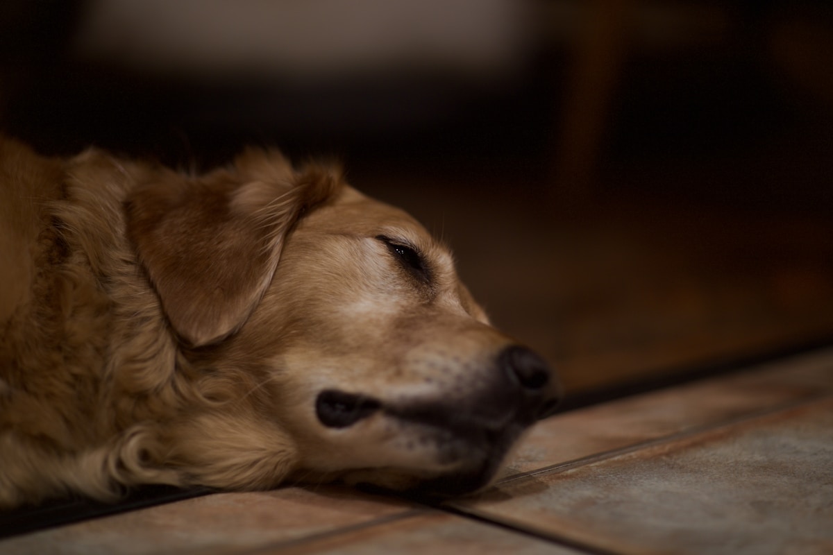
It is an all-too-familiar scene played out in millions of households every single day: your dog wakes up from a deep, peaceful afternoon nap on the living room rug, stretches out their front legs in a slow downward bow, and attempts to stand. As they take their first few steps toward the kitchen, you notice a distinct, hitching limp. One leg appears stiff, unyielding, and reluctant to bear weight, or their entire hindquarters sway awkwardly from side to side as they hobble forward. You watch with growing concern, your heart sinking as you wonder if they injured themselves during a morning walk. Yet, within thirty to sixty seconds of continuous movement, something remarkable happens: the limp begins to fade. By the time they reach the back door, their stride is smooth and symmetrical, and moments later, they are trotting around the backyard or chasing a favorite ball as if nothing was ever amiss.
Faced with this transient, warm-up pattern of lameness, many devoted dog owners talk themselves out of seeking veterinary care. They assume the dog simply slept in an awkward position, experienced a harmless muscle cramp, or had a limb fall asleep just as a human foot might after sitting cross-legged on the floor. In canine orthopedics, dogs do not experience transient limb paresthesia from sleeping postures in the way humans do. Limping that occurs exclusively or most severely upon rising from a period of prolonged rest is recognized by veterinary surgeons as the classic gelling phenomenon. Far from being an innocent sleeping cramp, it is one of the earliest, most pathognomonic clinical hallmarks of progressive articular cartilage degradation, chronic synovitis, and degenerative joint disease.
The tragedy of the joint gelling phenomenon is that its temporary nature lulls pet parents into a false sense of security. Because the dog walks it off within a minute, owners naturally assume the underlying issue is minor or self-resolving. Yet beneath the surface of the joint capsule, inflammatory cytokines, matrix metalloproteinases, and mechanical micro-frictional forces are actively eroding viable hyaline cartilage, setting the stage for permanent osteophyte proliferation and debilitating chronic pain. According to orthopedic consensus guidelines from the American Veterinary Medical Association (AVMA) and breed joint registries managed by the American Kennel Club (AKC), early therapeutic intervention during this initial gelling phase can preserve joint function and add years of comfortable, active life to your companion.
Bio-Rheology of the Diarthrodial Joint and the Gelling Phenomenon
To understand why a dog limps upon waking and then warms out of the stiffness, one must first explore the complex bio-rheology of a healthy diarthrodial (synovial) joint. A synovial joint: such as the canine hip, stifle (knee), hock, elbow, or shoulder: is an intricate biological bearing engineered for frictionless motion. The articulating bone surfaces are capped with smooth, glistening articular cartilage composed of specialized chondrocytes embedded within an extracellular matrix rich in Type II collagen fibers and large proteoglycan aggregates, predominantly aggrecan.
Encapsulating the entire joint is the fibrous joint capsule, lined internally by the delicate synovial membrane. This membrane is populated by specialized synoviocytes that synthesize and secrete synovial fluid: a thick, viscous biological lubricant composed primarily of high-molecular-weight hyaluronic acid, lubricin, and surface-active phospholipids. Under normal physiological conditions, synovial fluid has a friction coefficient lower than ice sliding on ice, allowing bone surfaces to glide over one another with near-zero mechanical resistance while delivering vital oxygen and nutrients to avascular chondrocytes.
Thixotropy and Non-Newtonian Fluid Mechanics
Synovial fluid is not a simple fluid; it is a thixotropic, non-Newtonian biological fluid whose viscosity changes dynamically in response to shear stress:
- Resting Molecular Entanglement: When a dog sleeps quietly during a two-hour nap, the shear rate within the joint drops to zero. In response, the long, branching chains of high-molecular-weight hyaluronic acid molecules interlock and aggregate, causing the fluid to thicken into a dense, semi-solid gelatinous state: a natural resting cushion designed to protect static joint structures.
- Shear-Thinning Transition: When the dog stands up and begins walking, mechanical shear forces break these non-covalent molecular bonds. The fluid rapidly shear-thins into a slippery, free-flowing liquid that coats the articular surfaces and allows frictionless gliding.
- Pathological Degradation in Osteoarthritis: In an inflamed, arthritic joint, synoviocytes and infiltrating inflammatory white blood cells release massive surges of pro-inflammatory cytokines, specifically Interleukin-1 beta (IL-1beta) and Tumor Necrosis Factor alpha (TNF-alpha). These cytokines trigger the release of matrix metalloproteinases (MMPs) and free radicals that fragment hyaluronic acid into short, low-molecular-weight chains.
- The Stiff Gel Trap: Consequently, osteoarthritic synovial fluid loses its elasticity. When the arthritic dog sleeps, the fragmented fluid thickens into an abnormally rigid, viscous sludge. Upon waking, taking the first few steps requires forcing dried, abrasive cartilage surfaces against this thick sludge, pinching exposed subchondral bone nociceptors and causing sharp pain until movement warms and thins the fluid.
Primary Orthopedic Pathologies Behind Post-Nap Limping
The vast majority of post-nap stiffness cases stem from structural developmental or degenerative orthopedic diseases affecting major weight-bearing joints.
1. Canine Hip Dysplasia and Coxofemoral Osteoarthritis
Canine Hip Dysplasia is the leading cause of hind-limb stiffness in medium, large, and giant-breed dogs, though it frequently affects smaller breeds as well:
- Joint Laxity: Congenital joint laxity prevents the femoral head from fitting snugly within the pelvic acetabulum (hip socket).
- Abnormal Biomechanical Shearing: As the dog walks and runs, abnormal micro-instability shears away the dorsal acetabular rim, stretching the richly innervated joint capsule.
- Post-Nap Presentation: Upon waking, the dog rises with slow, labored effort in the rear legs, displays a swaying or bunny-hopping gait, and drags the toes slightly before warming up into a steady trot.
2. Canine Elbow Dysplasia: The Forelimb Dilemma
Because dogs carry approximately sixty percent of their total body weight on their front legs, developmental elbow incongruity produces severe post-rest lameness:
- Fragmented Medial Coronoid Process (FMCP): Micro-fractures of the medial coronoid process of the ulna create loose bone fragments that act like a pebble inside a shoe, abrading humeral cartilage.
- Ununited Anconeal Process (UAP): Failure of the anconeal process growth plate to fuse leaves a large, unstable bony shelf inside the joint.
- Osteochondrosis Dissecans (OCD): Defective endochondral ossification leads to thickened, necrotic cartilage flaps on the humeral condyle.
- The Head-Bobbing Limp: Dogs with elbow dysplasia exhibit a distinct head-bob gait upon waking: their head dips downward when stepping on the sound forelimb and bobs sharply upward when weight is transferred to the painful, stiff limb.
3. Partial Tears of the Cranial Cruciate Ligament (CCL)
While an acute, complete rupture of the cranial cruciate ligament produces immediate, non-weight-bearing toe-touching lameness, many canine cruciate injuries begin as subtle, partial-thickness micro-tears:
- Chronic Synovial Effusion: In a partial tear, joint instability triggers continuous inflammatory fluid accumulation inside the stifle capsule. During a nap, this fluid pools under pressure.
- Rising Stiffness: When the dog rises, stretching the swollen joint capsule causes acute hesitation and limping for the first ten to fifteen strides before joint motion redistributes the fluid.
- The Medial Buttress: Over time, the body attempts to stabilize the loose knee by laying down dense fibrous scar tissue on the inside of the stifle, visible as a firm swelling known as a medial buttress.
Health certification guidelines from the Orthopedic Foundation for Animals (OFA) provide essential baseline data for evaluating structural soundness and hereditary predispositions.
Neurological Drivers Masquerading as Orthopedic Stiffness
Not every case of post-nap limping originates within a joint. Severe spinal cord and peripheral nerve compressions frequently mimic orthopedic morning stiffness.
Lumbosacral Stenosis (Cauda Equina Syndrome)
Lumbosacral stenosis is a progressive compressive neuropathy affecting the L7-S1 junction of the spine, predominantly striking large breeds like German Shepherds, Boxers, and Golden Retrievers:
- Sleeping Posture Flexion: When a dog curls up tightly into a ball to sleep, the lumbosacral spine is held in prolonged flexion, compressing inflamed nerve roots and reducing microvascular capillary perfusion to the sciatic nerve.
- Neurogenic Claudication: Upon rising, the dog experiences acute neurogenic numbness, tingling, and weakness in the hind legs. They rise with a low, tucked tail, scuff their toenails along hard floors, and wobble unsteadily for several paces until spinal extension restores blood flow to the nerves.
- Lumbosacral Palpation Pain: Direct downward pressure over the L7-S1 junction or hyperextension of the tail overhead elicits sharp cries of discomfort or defensive snapping.
Cervical Intervertebral Disc Disease (IVDD)
In smaller chondrodystrophic breeds (Dachshunds, French Bulldogs, Basset Hounds, Corgis), cervical disc herniation creates acute neck guarding upon rising. The dog lowers its head toward the floor, arches its upper back, trembles, and refuses to lift its head to greet you after waking, often limping on a front limb due to nerve root signature pain.
Soft Tissue and Muscular Injuries: Chronic Iliopsoas Strain
Active sporting dogs, hunting companions, and agility athletes frequently develop chronic micro-trauma in the major core muscles:
- Anatomy of the Iliopsoas Muscle: The iliopsoas is a powerful hip flexor composed of the psoas major and iliacus muscles, inserting onto the lesser trochanter of the femur.
- Traumatic Mechanism: Micro-tears occur when a dog slips on slick hardwood floors, lands awkwardly off agility obstacles, or makes explosive turns while chasing balls.
- The Short-Stride Post-Nap Limp: After resting in a shortened, contracted position during sleep, standing forces the inflamed iliopsoas tendon into sudden extension. The dog limps with a markedly shortened forward swing phase of the hind leg, avoiding full hip extension.
Clinical standards established by the American Animal Hospital Association (AAHA) emphasize the necessity of structured diagnostic evaluations and routine monitoring protocols.
Comprehensive Veterinary Clinical Orthopedic Examination
Accurately diagnosing the cause of post-nap limping requires a structured, multi-modal orthopedic and neurological workup performed by a qualified veterinary practitioner.
Static and Dynamic Gait Analysis
- Observation on Varied Surfaces: The clinician observes the dog rising from a seated or down position, walking, trotting, and turning in tight circles on both high-traction and smooth surfaces.
- Pressure-Sensitive Walkway Matometry: Advanced referral centers utilize computerized pressure walkways to objectively quantify the exact ground reaction force (peak vertical force and vertical impulse) distributed across each individual paw, detecting subclinical weight shifting long before it is visible to human eyes.
Orthopedic Palpation and Provocative Stress Maneuvers
- Cranial Drawer and Tibial Compression Tests: Palpates for abnormal forward translation of the tibia relative to the femur, confirming cranial cruciate ligament rupture.
- Ortolani Sign: Performed under heavy sedation to assess hip joint laxity and subluxation angle in dysplastic hips.
- Campbell’s Test: Evaluates collateral ligament integrity and joint incongruity in the canine elbow.
- Lumbosacral Tail Jack Maneuver: Elevating the tail base vertically while applying firm downward pressure over the L7-S1 junction isolates nerve root pain.
Diagnostic Imaging Modalities
- Digital Orthogonal Radiographs: High-resolution X-rays taken in orthogonal planes evaluate periarticular osteophyte formation, subchondral sclerosis, joint space narrowing, and bone remodeling.
- PennHIP Distraction Radiography: Measures the passive laxity of the hip joints using a specialized distractor device, calculating an objective Distraction Index (DI) that predicts the lifelong probability of osteoarthritis development.
- Musculoskeletal Computed Tomography (CT): The gold-standard imaging modality for canine elbow dysplasia, detecting fragmented medial coronoid processes and microscopic bone fissures that are completely invisible on standard plain X-rays.
- Magnetic Resonance Imaging (MRI): Indispensable for evaluating soft tissue structures, detecting intervertebral disc herniations, spinal cord compression, and acute iliopsoas muscle strains.
Modern Multimodal Medical Management of Canine Joint Pain
Modern veterinary rheumatology has transcended the outdated era of simply prescribing a generic pain pill and hoping for the best. Today, joint disease is managed through a sophisticated multimodal framework that addresses pain pathways from multiple biological angles.
1. Anti-NGF Monoclonal Antibody Therapy (Bedinvetmab / Librela)
The introduction of Bedinvetmab (marketed globally as Librela) represents the most significant breakthrough in veterinary arthritis management in over three decades:
- Mechanism of Action: Nerve Growth Factor (NGF) is a key biological mediator that drives osteoarthritis pain by binding to TrkA receptors on sensory nerve terminals, increasing pain signaling and promoting neurogenic inflammation. Bedinvetmab is a fully caninized monoclonal antibody that binds and neutralizes circulating NGF, preventing pain signals from reaching the central nervous system.
- Safety and Metabolism: Unlike NSAIDs, Bedinvetmab is eliminated via normal cellular protein catabolism through the reticuloendothelial system, bypassing hepatic metabolism and renal excretion. This makes it exceptionally safe for dogs with borderline kidney or liver values.
- Monthly Subcutaneous Dosing: Administered as a single monthly injection, it provides profound, continuous pain relief that drastically reduces or completely eliminates post-nap gelling.
2. Prostaglandin Receptor Antagonists (Grapiprant / Galliprant)
Traditional non-steroidal anti-inflammatory drugs (NSAIDs) inhibit cyclooxygenase enzymes (COX-1 and COX-2), reducing inflammatory prostaglandins but simultaneously risking gastric ulceration and renal hypoperfusion. Grapiprant represents a revolutionary piprant class of medication that specifically blocks the EP4 receptor: the primary prostaglandin receptor responsible for canine osteoarthritis pain: while sparing beneficial homeostatic prostaglandins throughout the gut and kidneys.
3. Disease-Modifying Osteoarthritis Drugs (DMOADs)
Polysulfated Glycosaminoglycans (Adequan Canine) and Pentosan Polysulfate (Cartrophen) are injectable therapeutics that actively slow joint degeneration:
- Enzyme Inhibition: DMOADs inhibit destructive enzymes (elastase, collagenase, and aggrecanases) within synovial fluid, halting the breakdown of hyaline cartilage.
- Synovial Fluid Regeneration: They stimulate synoviocytes to synthesize fresh, high-molecular-weight hyaluronic acid, restoring normal thixotropic lubrication and eliminating morning joint stickiness.
According to clinical pathology references published in the Merck Veterinary Manual, early identification of underlying physiological changes provides the highest probability of positive long-term therapeutic outcomes.
Transforming the Home Environment to Eliminate Joint Strain
Medical treatments must be supported by practical home modifications that protect healing joints from daily physical micro-trauma.
1. Orthopedic High-Density Memory Foam Bedding
Standard fiberfill dog beds compress completely under a dog’s weight, allowing elbows, hips, and hocks to press directly against hard flooring. Provide multi-layered orthopedic beds featuring medical-grade high-density support foam topped with gel-infused memory foam. This distributes weight evenly, eliminates pressure points, and maintains ambient warmth that keeps synovial fluid supple during naps.
2. High-Traction Floor Runners
Slick hardwood, laminate, and tile floors are treacherous for dogs with post-nap stiffness. When rising from sleep, a dog’s paws slip laterally, forcing unstable joints into sudden extension and tearing delicate micro-scar tissue. Place rubber-backed runner rugs, yoga mats, or high-traction carpets along primary household pathways, creating secure slip-free corridors.
3. Elevated Food and Water Bowls
Bending down to floor level to eat or drink forces a dog to shift sixty to seventy percent of their body weight onto their forelimbs and hyperextend the cervical spine. Raising feeding bowls to shoulder height allows your dog to maintain a neutral, pain-free spinal posture during meals.
4. Ramps and Vehicle Access Aids
Jumping into vehicles or off furniture exerts massive concussive impact forces on joints: up to four times the dog’s body weight upon landing. Install gentle, high-traction ramps to eliminate jumping entirely.
Weight Management: The Ultimate Biological Joint Shield
No surgical procedure, medication, or supplement can overcome the mechanical destruction caused by excess body weight. Adipose tissue (body fat) is not an inert storage depot; it is an active endocrine organ that pumps pro-inflammatory adipokines: including leptin, adiponectin, and interleukin-6: continuously into the bloodstream.
These circulating adipokines accelerate cartilage destruction and amplify nociceptive pain sensitivity. Clinical studies have conclusively proven that a modest weight reduction of just six to eight percent of total body weight results in profound, visible reductions in canine lameness and dramatically improves quality of life. Work closely with your veterinarian to calculate your dog’s exact Resting Energy Requirement (RER), replace high-calorie commercial biscuits with fresh green beans or carrot slices, and achieve a lean Body Condition Score (BCS) of 4 to 5 out of 9.
Regenerative Orthopedics: Platelet-Rich Plasma and Stem Cell Therapies
When conventional oral pharmaceuticals provide incomplete relief or when owners seek to minimize long-term NSAID usage, regenerative veterinary medicine offers cutting-edge biological tools that repair the internal joint environment.
Autologous Platelet-Rich Plasma (PRP) Therapy
Platelet-Rich Plasma is derived directly from your dog’s own blood through specialized gradient centrifugation, concentrating platelets up to five to seven times baseline physiological levels:
- Alpha Granule De-granulation: Once injected into the arthritic joint under sterile conditions, activated platelets degranulate, releasing potent anabolic growth factors: Transforming Growth Factor-beta (TGF-beta), Platelet-Derived Growth Factor (PDGF), and Vascular Endothelial Growth Factor (VEGF).
- Synovial Downregulation: These growth factors shut down catabolic inflammatory cytokines, suppress matrix metalloproteinases, and stimulate native synoviocytes to produce dense, high-molecular-weight hyaluronic acid.
- Clinical Longevity: A series of intra-articular PRP injections provides substantial reduction in post-nap gelling and joint effusion lasting between six and twelve months.
Adipose-Derived Mesenchymal Stem Cell (AD-MSC) Therapy
Mesenchymal stem cells harvested from your dog’s falciform adipose tissue possess profound immunomodulatory and paracrine signaling capabilities. When injected intra-articularly, MSCs home to areas of cartilage fibrillation, secreting bioactive trophic factors that prevent further chondrocyte apoptosis, reduce periarticular fibrosis, and restore smooth joint gliding.
Physical Rehabilitation Modalities: Photobiomodulation and Hydrotherapy
Restoring comfortable mobility requires non-invasive physical therapy modalities that relieve muscle tension and accelerate tissue repair without stressing vulnerable joints.
Class IV Photobiomodulation (Laser Therapy)
Deep tissue Class IV laser therapy delivers specific wavelengths of near-infrared light (typically 810 nm to 980 nm) deep into the joint capsule:
- Mitochondrial Chromophore Activation: Photons are absorbed by cytochrome c oxidase within the mitochondrial respiratory chain, stimulating rapid synthesis of adenosine triphosphate (ATP) and cellular reactive oxygen species scavenging.
- Rapid Analgesia and Vasodilation: Laser therapy promotes localized nitric oxide release, improving micro-capillary perfusion, reducing periarticular edema, and suppressing C-fiber nociceptive pain firing to eliminate the sharp hitch when rising from sleep.
Underwater Treadmill Hydrotherapy
Buoyancy is the ultimate friend of the arthritic dog. Water immersion up to the level of the greater trochanter bears up to sixty-two percent of your dog’s total body weight. Walking on an underwater treadmill allows the dog to flex and extend stiff joints through a full, natural range of motion with near-zero concussive impact, building supporting quadriceps and gluteal muscle mass while warm water keeps synovial fluid supple.
Targeted Joint Nutraceuticals: UC-II Collagen and Marine Fatty Acids
Internal nutritional support provides the biochemical precursors needed to maintain joint lubrication and suppress cartilage breakdown:
- Undenatured Type II Collagen (UC-II): Operates through a groundbreaking immunological mechanism known as oral tolerance. As tiny doses of native Type II collagen pass through the gut, they interact with Peyer’s patches in the gut-associated lymphoid tissue. This trains regulatory T-cells to migrate to inflamed joints and downregulate immune attacks against joint cartilage, significantly reducing morning stiffness.
- Purified Marine EPA and DHA: Cold-water marine fish oils rich in eicosapentaenoic acid incorporate directly into chondrocyte cell membranes, downregulating inflammatory COX-2 and aggrecanase enzymes (ADAMTS-4 and ADAMTS-5), halving cartilage breakdown rates.
- Green-Lipped Mussel Extract (Perna canaliculus): Rich in unique eicosatetraenoic acids (ETA) and natural glycosaminoglycans, green-lipped mussel provides rapid, natural inhibition of lipoxygenase and cyclooxygenase pathways without gastric irritation.
Surgical Interventions for Severe Structural Joint Instability
While multimodal medical management, weight control, and physical therapy form the cornerstone of arthritis care, severe mechanical joint instability or persistent bone-on-bone friction frequently requires definitive orthopedic surgical intervention to eliminate chronic post-rest pain.
1. Tibial Plateau Leveling Osteotomy (TPLO) for CCL Rupture
When cranial cruciate ligament micro-tears progress to partial or full mechanical instability, medical management alone cannot prevent severe secondary osteoarthritis. The TPLO procedure surgically alters the biomechanics of the stifle joint:
- Neutralizing Cranial Tibial Thrust: A semi-circular radial cut is made through the proximal tibia, rotating the tibial plateau to reduce its slope from a steep twenty-five to thirty degrees down to a stable five to six degrees.
- Eliminating the Need for a Ligament: By leveling the plateau, the femur no longer slides backward off the tibia during weight-bearing, converting shearing forces into pure axial compression and immediately stabilizing the knee.
- Rapid Bone Healing: Secured with a rigid medical-grade titanium compression plate, bone union occurs within eight to ten weeks, permanently stopping post-nap joint gelling in the affected limb.
2. Total Hip Replacement (THR) and Femoral Head Ostectomy (FHO)
For canine patients suffering from intractable hip dysplasia where cartilage is completely worn away, two primary surgical pathways provide dramatic pain relief:
- Total Hip Replacement (THR): The gold-standard biomechanical procedure that completely replaces the diseased acetabulum and femoral head with state-of-the-art prosthetic components (a high-density polyethylene cup and titanium femoral stem). THR completely eliminates arthritic friction, restoring normal, pain-free joint biomechanics for the remainder of the dog’s natural lifespan.
- Femoral Head Ostectomy (FHO): A salvage procedure predominantly utilized in small to medium dogs or when financial constraints preclude THR. The femoral head and neck are surgically excised, allowing the surrounding gluteal muscles and a dense pseudoarthrosis (fibrous scar joint) to support the limb without painful bone-on-bone contact.
3. Arthroscopic Debridement for Elbow Dysplasia
Minimally invasive arthroscopy allows orthopedic surgeons to insert microscopic fiber-optic cameras and precision motorized micro-shavers into the elbow joint:
- Fragment Removal: Surgeons excise loose, fragmented medial coronoid processes (FMCP) and smooth out fibrillated articular cartilage flaps with sub-millimeter precision.
- Microfracture Chondroplasty: Tiny micro-punctures are made into the exposed subchondral bone, stimulating mesenchymal stem cells from the bone marrow to form a protective fibrocartilage cap over bare bone areas.
Thermal Therapy at Home: Moist Heat Compresses and Controlled Warming
One of the most effective, immediate non-pharmacological methods to alleviate post-nap joint gelling is targeted thermal therapy. Applying controlled, gentle warmth to stiff hips, stifles, or elbows before a dog rises delivers immediate physiological benefits:
Microvascular Vasodilation and Lubrication Flow
When a joint rests in a cool environment, local blood vessels constrict, and synovial fluid thickens. Applying warmth for ten to fifteen minutes produces immediate biological changes:
- Increased Micro-Capillary Perfusion: Radiant heat dilates local blood vessels, delivering fresh oxygenated blood to chronically tight periarticular muscles and fascia.
- Accelerated Thixotropic Shear-Thinning: Warmth raises intra-articular temperature, lowering the resting viscosity of synovial fluid before the dog even takes its first step.
- Nociceptive Pain Gate Closing: Thermal receptors stimulate large-diameter sensory nerve fibers, which functionally close the pain gate in the dorsal horn of the spinal cord, blocking sharp aching signals.
Safe Application Guidelines
Use a commercial moist-heat clay pack or a warm damp towel wrapped inside a thin cotton pillowcase. Warm it to a comfortable lukewarm temperature (test it thoroughly against your own inner wrist first). Never use high-voltage human electric heating pads, as canines cannot move away quickly if the device overheats, creating serious burn risks. Gently drape the warm pack over the stiff joint for ten to twelve minutes as your dog begins to stir from sleep.
Clinical Diagnostic Frameworks: Biomarker Analysis and Preventative Veterinary Care
Navigating canine health challenges requires a systematic diagnostic methodology grounded in evidence-based veterinary pathology. Whether evaluating insidious systemic complaints such as mild polydipsia, post-rest lameness, or subtle ocular alterations, clinical specialists emphasize the necessity of combining objective laboratory biomarkers with detailed longitudinal symptom tracking. Routine baseline screening – encompassing complete blood counts, comprehensive biochemical profiles, high-resolution digital imaging, and specific serological antibody assays – enables veterinary teams to intercept metabolic and degenerative disorders during subclinical stages.
Cellular Pathology: Microvascular Perfusion and Endothelial Function in Canine Disease
Systemic illness in canines frequently manifests through alterations in microvascular capillary perfusion and endothelial barrier permeability. Inflammatory cytokines released during infectious or autoimmune states stimulate endothelial cell activation, promoting localized vascular stasis, plasma extravasation into interstitial spaces, and micro-thrombosis. Monitoring capillary refill time, serum lactate levels, and base excess provides clinicians with real-time metrics of tissue oxygenation and hemodynamic stability, guiding targeted fluid therapy and pharmacological interventions before organ dysfunction becomes irreversible.
Immunological Homeostasis: Managing Chronic Enteropathies and Systemic Inflammation
The gut-associated lymphoid tissue (GALT) plays a central role in maintaining systemic immune tolerance while protecting against enteric pathogens. Disruptions in mucosal tight junctions (colloquially termed intestinal permeability) permit dietary antigens and bacterial lipopolysaccharides to enter the lamina propria, triggering chronic low-grade inflammation that can manifest as intermittent gastrointestinal borborygmi, selective appetite loss, or distant dermatological flare-ups. Implementing targeted hypoallergenic diets, mucosal cytoprotectants, and scientifically validated synbiotic therapy restores mucosal barrier integrity and stabilizes immune equilibrium.
Geriatric Canine Wellness: Comprehensive Palliative Strategies and Quality of Life
Optimizing senior canine longevity requires a multi-modal wellness approach that addresses cognitive preservation, joint comfort, and metabolic stability. Incorporating omega-3 polyunsaturated fatty acids (EPA and DHA), multimodal analgesia protocols (including modern anti-NGF monoclonal antibodies), and gentle low-impact physical exercise maintains lean muscle mass and cognitive engagement. Regular veterinary health evaluations every six months ensure that age-related degenerative changes are managed proactively, preserving dignity and comfort throughout the golden years.
Veterinary Orthopedic Progression Monitoring and Multimodal Pain Audits
Maintaining long-term wellness for companion dogs requires an active, ongoing partnership between dedicated guardians and qualified veterinary professionals. Establishing regular six-month wellness examinations, conducting routine diagnostic laboratory panels, and maintaining longitudinal records of behavioral patterns and dietary responses provides clinicians with essential baseline metrics. By adopting an evidence-based preventative mindset and addressing subtle environmental and physical changes promptly, pet parents ensure their canine companions enjoy optimal vitality, emotional comfort, and flourishing health throughout every developmental life stage.
Comparative Diagnostic Matrix: Causes of Post-Nap Lameness
To assist owners and veterinarians in identifying the source of post-nap stiffness, the table below compares the hallmark clinical features, physical findings, and interventions for primary conditions:
| Underlying Pathology | Typical Post-Nap Presentation | Age and Breed Profile | Key Physical Exam Finding | First-Line Therapeutic Intervention |
|---|---|---|---|---|
| Hip Dysplasia / OA | Labored rear rise, swaying hindquarters | Medium to large breeds, adult to senior | Pain on hip extension, reduced abduction range | Anti-NGF monoclonal antibodies, weight control |
| Elbow Dysplasia (FMCP) | Head-bobbing forelimb limp, outward paw rotation | Labradors, Bernese, Shepherds, 1 to 5 years | Joint effusion, pain on terminal elbow flexion | Arthroscopic fragment removal, PSGAG therapy |
| Partial CCL Micro-Tear | Hesitant toe-touching, stiff stifle for 20 paces | Any breed, overweight dogs, 3 to 8 years | Positive cranial drawer sign, medial buttress | Surgical stabilization (TPLO) or strict rest |
| Lumbosacral Stenosis | Wobbly hind rise, scuffing toenails, low tail | German Shepherds, Boxers, 6+ years | Pain on L7-S1 dorsal palpation and tail jack | Spinal anti-inflammatories, epidural injections |
| Chronic Iliopsoas Strain | Shortened hind stride, reluctant to extend hip | Agility dogs, hunting dogs, active adults | Direct pain on internal pelvic tendon palpation | Regenerative shockwave therapy, hydrotherapy |
| Immune-Mediated Polyarthritis | Severe all-limb stiffness, walking on eggshells | Any breed, young adult to middle-aged | Swollen warm carpi and hocks, fluctuating fever | Immunosuppressive corticosteroid protocol |
Frequently Asked Questions About Dog Limping After a Nap
Why does my dog limp right after waking up but walk normally a minute later?
This is known as the gelling phenomenon. During sleep, fragmented synovial fluid in an arthritic joint thickens into a stiff gel, causing frictional resistance and sharp pain on the first few steps. As the dog walks, movement warms and shear-thins the fluid back into a liquid state, temporarily relieving the pain until the next rest period.
Is post-nap limping simply caused by a dog’s leg falling asleep?
No. Dogs do not experience transient limb paresthesia (pins and needles) from sleeping postures like humans do. Limping upon rising is almost always an early clinical sign of joint inflammation, cartilage erosion, osteoarthritis, or spinal nerve root compression.
Can young dogs develop post-nap stiffness?
Yes. Young dogs between six and eighteen months of age can display post-nap stiffness due to developmental orthopedic diseases, including hip dysplasia, elbow dysplasia (fragmented medial coronoid process), panosteitis (growing pains), or osteochondritis dissecans (OCD).
What is the difference between hip dysplasia and elbow dysplasia in how a dog limps?
Hip dysplasia causes hind-limb stiffness, characterized by difficulty rising, a swaying rear end, and a bunny-hopping gait on stairs. Elbow dysplasia causes forelimb stiffness, marked by a distinct head-bobbing gait where the head drops on the good leg and lifts on the painful limb.
Should I let my dog run and play fetch if they limp after naps?
You should avoid high-impact, explosive activities like chasing tennis balls, jumping, and rough agility play, as repetitive concussive impacts accelerate cartilage degradation. Instead, transition to controlled, low-impact exercise such as steady leash walks on flat ground and swimming.
Can cold or damp weather make post-nap limping worse?
Yes. Drops in barometric pressure cause subtle expansion of inflamed joint capsules, while cold temperatures decrease ambient blood flow and increase the resting viscosity of synovial fluid, intensifying morning stiffness and joint discomfort.
What is Bedinvetmab (Librela) and how does it help?
Bedinvetmab is a fully caninized monthly injectable monoclonal antibody that binds and neutralizes Nerve Growth Factor (NGF). By preventing NGF from binding to pain receptors, it safely halts arthritis pain signaling without stressing the kidneys or liver.
Can giving human Tylenol or Ibuprofen relieve my dog’s morning limp?
NEVER give human pain medications like Tylenol (acetaminophen), Advil (ibuprofen), or Aleve (naproxen) to a dog. They are highly toxic to canines, causing life-threatening gastric ulceration, intestinal perforation, acute liver necrosis, and fatal kidney failure.
How does weight loss help reduce joint stiffness?
Losing weight reduces the mechanical force exerted across bearing joints with every step. Additionally, body fat secretes inflammatory adipokines into the bloodstream; losing fat eliminates systemic inflammatory signals that drive joint pain.
What are the benefits of an orthopedic memory foam bed?
High-density memory foam distributes your dog’s body weight evenly, preventing bony joints (elbows, hips, hocks) from pressing against hard floors. It also insulates joints against cold drafts, keeping synovial fluid warmer and more fluid during sleep.
Restoring Effortless and Joyful Mobility to Your Companion
Watching your loyal companion struggle to rise from their bed: their limbs trembling slightly, their stride stiff and tentative: can be a heart-wrenching experience for any pet parent. Yet, recognizing the gelling phenomenon for what it truly is: an early physiological alarm bell rather than an innocent sleeping cramp: empowers you to take decisive, compassionate action before irreversible cartilage destruction takes hold.
Armed with an understanding of synovial bio-rheology, thixotropic fluid mechanics, and the crucial differences between developmental joint dysplasia, cruciate tears, and neurological compressions, you are prepared to be your dog’s greatest health advocate. You now know that a limp that fades after twenty steps is not a minor quirk to be ignored, but an urgent invitation to partner with your veterinarian. Through precise orthopedic diagnostics, cutting-edge monoclonal antibody therapies, targeted DMOAD injections, and modern anti-inflammatory medications, you can dissolve joint stiffness at its molecular source.
Pair these clinical interventions with the transformative power of home care: providing luxurious orthopedic memory foam beds, laying down high-traction floor runners, and maintaining a lean, athletic body condition that unburdens overworked joints. When you build this comprehensive circle of orthopedic care, the results are deeply rewarding: your dog rises from every nap with ease and comfort, their eyes bright, their tail wagging, ready to share every joyful adventure by your side for all the happy years ahead.

Leave a Comment